Diagram Of Liver Cell : Rat Liver Cell Biological Activity Cell Life - Ƽ store vitamins and minerals;
byAdmin•
0
Diagram Of Liver Cell : Rat Liver Cell Biological Activity Cell Life - Ƽ store vitamins and minerals;. Expression of liver specific proteins decreases with time in culture, but is reactivated by growing the cells in serum free medium. Связки печени ligaments of the liver. Hepatocytes are polygonal epithelial cells with abundant eosinophilic, granular cytoplasm and large, centrally located round nuclei. Whatever an organism does for survival it does for the survival of its cells. 12.08.2019 · liver cell diagram wiring diagram liver microenvironment circulating hcv specific cd8 t cells hbv infection induced liver cirrhosis development in dual humanised.
The human liver is an essential multifunctional organ. 2.3.1 draw and label a diagram of the ultrastructure of a liver cell as an example of an animal cell. Hepatocyte nuclei often contain a prominent nucleolus. It may be also regarded as the basic unit of biological activity. Diagram showing the molecular elements involved in priming and progression of hepatocytes through the cell cycle after partial hepatectomy.
Cytokines In Adipose Derived Mesenchymal Stem Cells Promote The Healing Of Liver Disease from f6publishing.blob.core.windows.net It may be also regarded as the basic unit of biological activity. It should be large, clear and with specific labels. The liver performs many essential functions related to digestion, metabolism, immunity, and the storage of nutrients within the body. Pharmacotoxicological studies and for the investigation of. | human cell structure, animal cell project, animal cell. The cell is the structural and functional unit of life. The liver parenchyma is primarily comprised of hepatocytes. Animal liver cell diagram ~ diagram.
An in vitro model for.
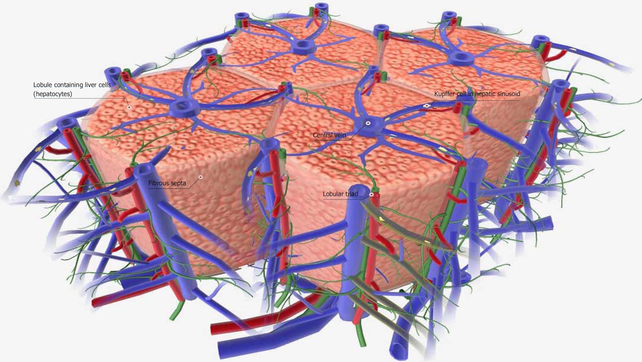
No previous treatment for liver cell damage. The liver parenchyma is primarily comprised of hepatocytes. Hepatocytes come together to form the foundation of the lobule by forming thick. The liver is an accessory digestive organ that produces bile, an alkaline fluid containing cholesterol histology, the study of microscopic anatomy, shows two major types of liver cell: Documents similar to liver pathophysiology and schematic diagram. The cell is the structural and functional unit of life. The liver performs many essential functions related to digestion, metabolism, immunity, and the storage of nutrients within the body. Hepatocyte nuclei often contain a prominent nucleolus. Medical labeled diagram with all kind cells. However, the cellular composition of the liver remains poorly understood. Blood flows through the liver. Another type of liver cell is the endothelial cells. Smartdraw includes 1000s of professional healthcare and anatomy chart templates that you can modify and make your own.
Whatever an organism does for survival it does for the survival of its cells. No previous treatment for liver cell damage. Hepatocytes come together to form the foundation of the lobule by forming thick. Two diagrams of liver structure removed for copyright reasons. 7710x4991 liver cell diagram liver histology labpedia.
Schematic Diagram Of The Normal Liver At The Microscopic Level The Download Scientific Diagram from www.researchgate.net The incidence of liver diseases is rising and there are limited treatment options. Form specific compounds such as coagulation factors and. Связки печени ligaments of the liver. The bandpass can be varied in the following ways: Create healthcare diagrams like this example called liver cells in minutes with smartdraw. The cells of the living kingdom may be divided into two categories: Signs and symptoms of liver disease include abdominal pain, jaundice, nausea, and weakness. Another type of liver cell is the endothelial cells.
However, the cellular composition of the liver remains poorly understood.
2.3.2 annotate the diagram from 2.3.1 with the functions of each named structure. Liver cells express mscca (bear, 1990) and previous studies had shown that osmotic swelling of epithelial cells activates an mscca‐dependent figure 5.7. Causes, treatment, and life expectancy vary. The cell is the structural and functional unit of life. Smartdraw includes 1000s of professional healthcare and anatomy chart templates that you can modify and make your own. Medical labeled diagram with all kind cells. It should be large, clear and with specific labels. No previous treatment for liver cell damage. Ƽ intricately involved in carbohydrate, fat, and protein metabolism. These functions make the liver a vital organ without which the tissues of the body would quickly die from lack of energy and nutrients. An in vitro model for. Связки печени ligaments of the liver. Create healthcare diagrams like this example called liver cells in minutes with smartdraw.
It should be large, clear and with specific labels. Pharmacotoxicological studies and for the investigation of. Learn vocabulary, terms and more with flashcards, games and other study tools. Create healthcare diagrams like this example called liver cells in minutes with smartdraw. Control of liver cell fate decision by a gradient of tgf beta signaling modulated by onecut transcription factors.
Cell Diagram from animal.memozee.com Diagram showing the molecular elements involved in priming and progression of hepatocytes through the cell cycle after partial hepatectomy. Liver medicine refers to all diagnostic and treatment strategies of diseases and conditions that cause liver failure directly or indirectly. Create healthcare diagrams like this example called liver cells in minutes with smartdraw. Medical labeled diagram with all kind cells. Two diagrams of liver structure removed for copyright reasons. Causes, treatment, and life expectancy vary. Expression of liver specific proteins decreases with time in culture, but is reactivated by growing the cells in serum free medium. Example of blood, neurons, cardiac, bone, intestinal, epithelial, fat, liver and.
Control of liver cell fate decision by a gradient of tgf beta signaling modulated by onecut transcription factors.
An in vitro model for. It should be large, clear and with specific labels. Human anatomy detailed diagram of various human organs liver, heart, kidneys, lungs, colon, intestine, stomach, brains, etc can be used in. Binucleated hepatocytes (= containing two nuclei). Example of blood, neurons, cardiac, bone, intestinal, epithelial, fat, liver and. A fixed, narrow bandpass, which is centred round a middle frequency. Learn vocabulary, terms and more with flashcards, games and other study tools. Hepatocytes are polygonal epithelial cells with abundant eosinophilic, granular cytoplasm and large, centrally located round nuclei. Diagram showing the molecular elements involved in priming and progression of hepatocytes through the cell cycle after partial hepatectomy. However, the cellular composition of the liver remains poorly understood. Lifestyle changes may slow the progression of some types of liver disease. The incidence of liver diseases is rising and there are limited treatment options. The cell is the structural and functional unit of life.
Ƽ store vitamins and minerals; diagram of liver. Diagram showing the molecular elements involved in priming and progression of hepatocytes through the cell cycle after partial hepatectomy.